
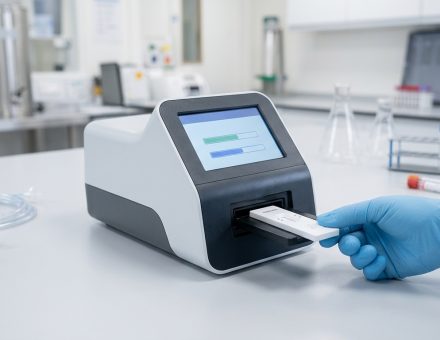
Medical imaging isn’t one market. It’s at least six, and each has its own optical priorities: diagnostics needs flat, repeatable fields; endoscopy needs small diameters and wide views; surgical systems need contrast and depth; veterinary devices need durability at a lower price point; life-science instruments need precision around the clock; and contract manufacturers need a partner who can scale. Across all six, the same pattern works: start with a proven off-the-shelf lens to validate the concept quickly, then move to a custom design and an active-aligned camera module when the requirements call for it. The lenses named here are examples, not prescriptions. The right solution comes out of a detailed conversation about your device.
Medical Imaging Optics: From Off-the-Shelf Lenses to Custom Camera Modules in Six Applications
In the medical market, we see six higher-level application segments: diagnostics, endoscopy, surgical, veterinary, life sciences, and contract manufacturing. Each one brings a different first question to our engineering team.





Diagnostic
Endoscopy
Surgical
Veterinary
Life Science
Contract MFG.
A diagnostic reader team asks about distortion at a 20 mm working distance. An endoscope team asks how small the optical stack can get. A surgical robotics group asks about veiling glare under bright LED illumination. Same lens portfolio. Very different conversations.
This article walks through all six. For each one, we describe the imaging challenge we typically see, where an off-the-shelf Sunex lens can get a program moving, and where a custom design usually takes over. We close with camera modules and active alignment, because for many medical devices that is where image quality is finally won or lost.
Please note: the examples are examples only. Many other solutions exist.
The applications and part numbers in this article illustrate typical starting points. They are not a recommendation for your specific device. Optical requirements in medical imaging depend on the sensor, working distance, illumination, regulatory class, reprocessing method, and volume, so we always work through those details with each customer before we suggest a lens or a design path.
Why does almost every medical imaging program start in the same place?
Because the first goal is nearly always the same: get real images from the real sensor as early as possible. Two things we offer make that practical, and they work best together.
A large off-the-shelf lens portfolio for fast, low-risk starts
Our lens portfolio covers production lenses in M7, M8, M10, M12, M14, and M16 mounts, with focal lengths from under 1 mm (miniature fisheye) to about 20 mm (narrow field of view), and filter options that include IR-cut, no-IR, dual-band day/night, and NIR bandpass versions. Sample quantities can be ordered online, so a feasibility camera can be imaging within days rather than months.
If you’re building a feasibility prototype for a Class II device, an off-the-shelf lens lets your team produce real images for design-input discussions before anyone commits to optical tooling. That alone often settles arguments about field of view and resolution that would otherwise run for weeks.
More than 30 years of custom design and high-volume manufacturing
When an off-the-shelf lens can’t meet the envelope, the working distance, or the image quality target, our optical design team takes over. Over three decades, we’ve designed and built all-glass, all-plastic, and hybrid glass-plastic lenses, including proprietary approaches such as Tailored Distortion® (controlling how the field maps onto the sensor), NoGhost™ (suppressing ghost reflections at the design stage), SuperFisheye™ wide-angle optics, and the DXM™ dual-lens, single-sensor stereo platform. The same factories that produce automotive-grade volumes build the custom designs, so a prototype design does not have to be re-engineered when it moves to production.
Off-the-shelf vs. custom: how the two paths compare
| Criterion | Off-the-shelf lens | Custom lens design |
|---|---|---|
| Time to first images | Days (order samples online) | Weeks to months (design, prototype, test) |
| Upfront cost | Sample price only, no NRE | Design NRE plus tooling for molded or ground parts |
| Optimized for your sensor and working distance | Close, rarely exact | Yes, by design |
| Mechanical fit in tight envelopes | Limited to standard barrels and mounts | Shaped around your housing |
| Unit cost at volume | Fixed list pricing | Typically lower once tooling is amortized |
| Best for | Feasibility, early clinical prototypes, low-volume devices | Production devices with defined requirements and volume |
One nuance we discuss with almost every medical customer: volume changes the right design form. A molded aspheric element can replace two spherical elements, but the mold has to be paid for. For low-volume medical programs, an all-spherical or glass design can be the better economic choice even if it carries one more element.
An off-the-shelf lens is not a shortcut through verification
Whichever path you choose, the device manufacturer’s design controls still apply. We supply the optical data; your team still has to verify the lens in your system, under your conditions..
What does a diagnostic reader really need from its lens?
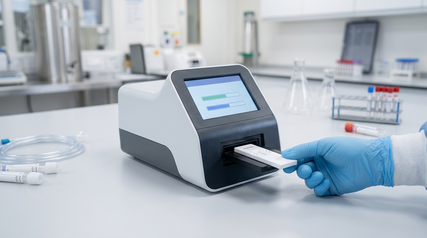
Diagnostic imaging covers point-of-care readers, lateral-flow and cartridge readers, assay and sample imaging, and screening cameras. Most of these image a test strip, cartridge window, or sample well at a short, fixed distance, and then measure something. That last word matters. The lens isn’t producing a picture for a person; it’s producing data for an algorithm.
That shifts the priorities:
- Low distortion and a flat field, so measurement regions land in the same pixels on every unit.
- Uniform relative illumination, so intensity readings aren’t biased toward the center of the field.
- Spectral control, with filters matched to the colorimetric or fluorescent markers being read.
- Unit-to-unit repeatability, because the algorithm is typically calibrated once for the whole product line.
Good off-the-shelf starting points are low-distortion M12 lenses such as the DSL949A (1/3″ format, EFL 3.4 mm, F/2.0) or the DSL355A (up to 1/2.5″, EFL 4.16 mm, F/2.8). Where reader height is tight, low-profile glass lenses like the DSL829L (M7 mount, EFL 5.9 mm, F/3.0) keep the optical stack short.
Here’s the limitation. Most off-the-shelf lenses are optimized for longer object distances. Refocusing them to a 15 to 30 mm working distance usually works for feasibility, but we check the MTF at the actual conjugate before anyone relies on it. A production reader often justifies a finite-conjugate custom design that is optimized at exactly that distance, with tolerance to cartridge seating height built in.
CUSTOMER SCENARIO
If your reader quantifies line intensity on a lateral-flow strip, a relative illumination falloff of a few percent from center to edge can shift results along the strip. Calibration can absorb that, but only if the falloff is consistent from unit to unit. That’s a lens-and-assembly question, not just a lens question.
How small can endoscope optics get before you need a custom design?
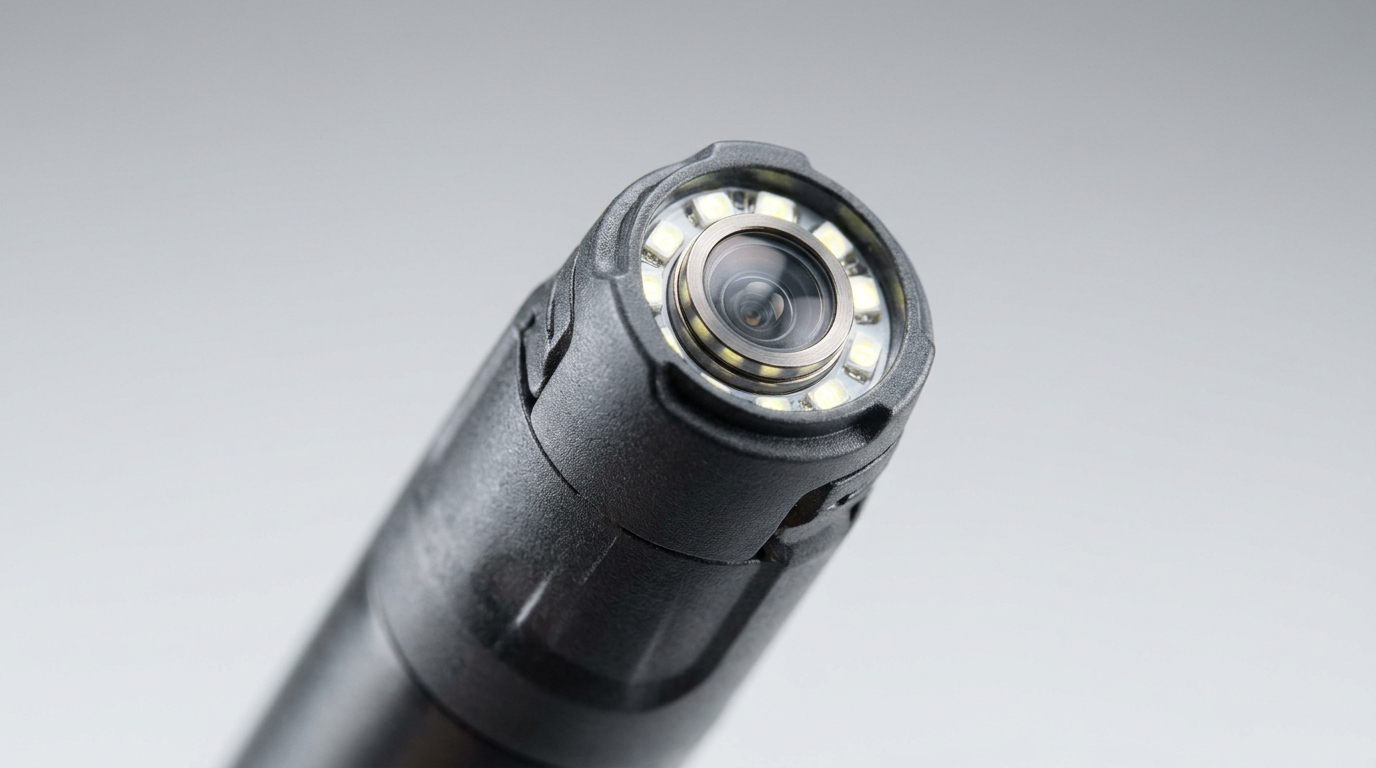
Endoscopy combines the hardest constraints in one package: a small diameter, a wide field of view, a depth of field that runs from a few millimeters to several centimeters, small-pixel chip-on-tip sensors, and a housing that has to survive reprocessing or be cheap enough to discard after a single use.
Let’s be direct about the boundary. The smallest threaded lenses in our portfolio use M7 and M8 mounts. They suit larger-diameter scopes, visualization handpieces, and proof-of-concept rigs well. For distal tips under roughly 8 mm, an off-the-shelf lens won’t fit, and the program needs a custom miniature design from the start.
Within that boundary, the lens portfolio gets early work moving:
- DSL235D: low-profile megapixel lens for 1/3″ sensors, M8x0.35 metal barrel, EFL 3.02 mm, F/2.2 or F/3.2, with a lens cap that includes an O-ring gland for sealing.
- DSL180A: miniature Tailored Distortion fisheye, EFL 0.97 mm, F/2.0, for very wide fields where more pixels should stay on the center of the scene.
- DSL829L: M7 low-profile glass lens when length matters more than field of view.
Custom endoscope work usually centers on three decisions: the f-number (which trades light against depth of field in a fixed-focus design), the front window and how it is sealed, and how the distal module is assembled and aligned. We cover that last point in the active alignment section below.
CUSTOMER SCENARIO
For a single-use endoscope, the optics can be one of the largest cost lines in a device that is discarded after one procedure. That moves the design conversation toward molded plastic elements and assembly steps that scale, which is exactly where our high-volume manufacturing experience pays off.
Why do surgical visualization systems live or die on stray light and depth?
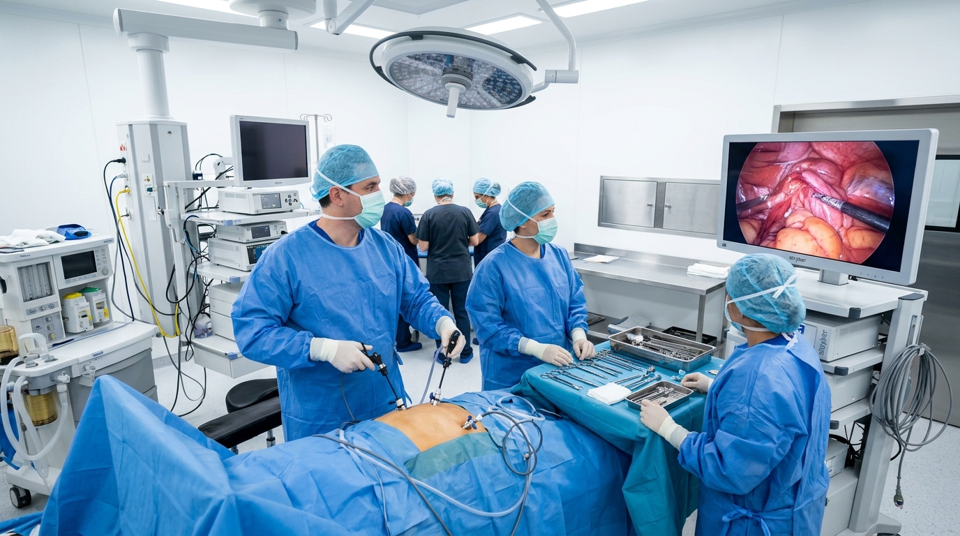
Surgical imaging, whether laparoscopic, open-field, or robotic, happens under intense illumination, with wet tissue and polished instruments throwing specular reflections back toward the lens. Every multi-bounce path inside the lens can become a ghost or a veil of flare, right where the surgeon is looking. Add HDR sensors, strict color fidelity, and growing use of near-infrared fluorescence, and the optical requirements stack up quickly.
Two Sunex capabilities map directly onto those problems.
NoGhost™ for contrast under bright light
NoGhost designs analyze and suppress ghost paths during optical design instead of trying to mask them later in software. Off-the-shelf examples include the DSL977A (1/3″ HDR sensors, EFL 7.7 mm, F/1.8) and the DSL957A (1/2.5″ HDR sensors, EFL 7.7 mm, F/1.8). For systems that image in both the visible and NIR bands, dual-band lenses such as the DSL893A RGBIR design show how one lens can serve both, although fluorescence bands usually call for custom coatings matched to the specific dye.
DXM™ for stereo depth from one sensor
DXM places two optical paths onto a single image sensor. The result is a stereo pair that is inherently synchronized, shares one readout, and lives in one module, which simplifies 3D visualization and depth measurement for robotic platforms.
Where DXM is not the perfect fit
Because both views share one sensor, the stereo baseline is limited by the sensor’s width. That suits short working distances well. If your system needs precise depth at longer distances, DXM with relay optics, or a two-camera arrangement with a wider baseline may serve you better, and we’ll tell you so.
Can veterinary imaging reuse human-medical optics, and where should it differ?
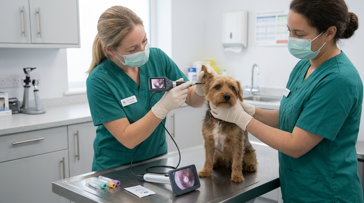
In many cases, yes. Veterinary endoscopy, dental imaging, and examination cameras share most of their optical physics with their human counterparts. Three differences change the design conversation, though.
- Price points are lower, so large NRE budgets are rare, and off-the-shelf lenses carry more of the load.
- Patient size ranges from small animals to horses. An equine gastroscope, for example, can run 3 meters or more at a diameter of around 13 mm.
- Use conditions are harder: barns, mobile clinics, washdown, and drops are part of normal life for the equipment.
The regulatory path is also different. In the US, the FDA does not require 510(k) or premarket approval submissions for veterinary devices, which typically shortens the route from prototype to product. That makes an off-the-shelf-first approach especially attractive.
For harsh environments, the DSL958R (EFL 4.6 mm, F/1.7, M12) is front-side sealed to IP6K9K, and the DSL383G SuperFisheye lens combines NoGhost and Tailored Distortion with an optional HP3 hydrophobic coating that makes the front surface easier to keep clean. Where a human-medical design already exists, a derivative with lower-cost materials or a simplified mechanical design is often the efficient path.
CUSTOMER SCENARIO
If you’re adapting a human-medical scope platform for a veterinary line, ask early which requirements can be relaxed. Dropping an autoclave requirement or widening a tolerance can remove cost without touching the image quality that the clinician actually sees.
What makes life-science and lab-automation optics different from a microscope?
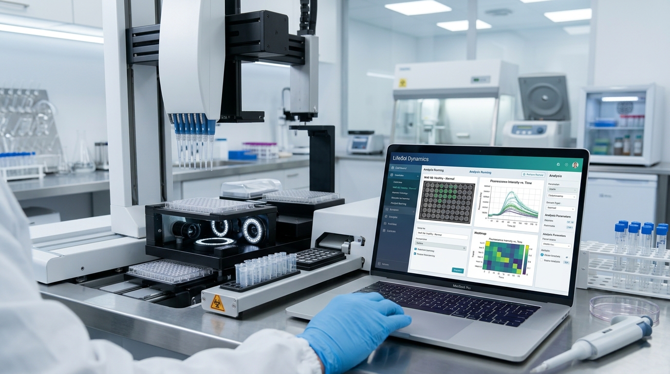
Life-science instruments image microplates, tubes, colonies, cells, and labels, often around the clock and often with several cameras inside one instrument. The microscope is still there for detailed work. Around it, a growing number of embedded cameras handle plate imaging, sample tracking, liquid-level checks, and barcode reading.
These cameras need measurement accuracy over a large field, consistent performance between the cameras in one instrument, and stable focus inside enclosures that often run warm, such as incubators held at 37 °C.
From the lens portfolio, the DSL958G family (EFL 4.6 mm, M12) is a useful low-distortion reference because it comes in F/1.7, F/2.8, and F/4.0 versions. That lets an instrument team test the trade between light gathering and depth of field with the same field of view. For higher magnification at a distance, narrow-field glass lenses such as the DSL330A (EFL 12 mm, M14) and DSL331A (EFL 19.8 mm, M16) are good starting points.
Custom work in this segment often involves near-telecentric designs (so that magnification doesn’t change with small height variations), athermalized designs for stable focus across temperature, and matched lens sets for multi-camera instruments.
CUSTOMER SCENARIO
If an instrument images a full SBS-format microplate (about 128 x 85 mm) in one frame, distortion at the corners decides whether well-position software needs per-unit calibration. Tailored Distortion can be designed so the well grid maps cleanly onto the sensor.
How should medical OEMs and contract manufacturers engage an optics partner?
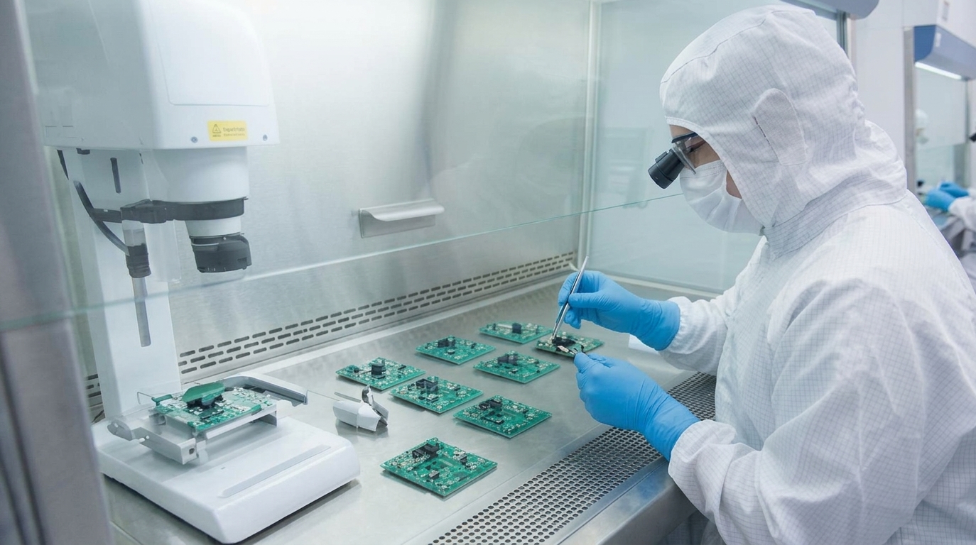
Contract manufacturers build to someone else’s design and carry the supply-chain risk for it. In our experience, the optics often arrive as a black-box part number, and the questions that matter most (lifecycle, change control, inspection data) come up late. They are much easier to answer early.
We support contract manufacturers and OEMs in three ways: off-the-shelf lenses as fast references for quoting and design-for-manufacture reviews, custom designs built to the customer’s specification, and high-volume production of lenses and camera modules. Lens holders and thread adapters (M7, M8, M10 and M12) from the same portfolio make it simple to mount a candidate lens on an existing board during evaluation.
Questions to settle with any optics supplier before a medical program ramps
| Topic | What to agree on early |
|---|---|
| Product lifecycle | Expected availability window, last-time-buy process, and redesign notice period |
| Change control | Which changes (materials, coatings, sites, processes) trigger notification |
| Inspection data | Which parameters are measured per lot (for example MTF, focal length, centering) |
| Documentation | Drawings, material declarations, and test reports your quality system needs |
| Cleanliness and packaging | Particle and handling requirements for assembly in controlled environments |
Who holds regulatory responsibility
We design and build optics and camera modules. Regulatory responsibility for the finished medical device stays with the device manufacturer. Our role is to supply the optical data, documentation, and change discipline your quality system requires, and we discuss those requirements in detail at the start of a program.
Why do active-aligned camera modules give medical devices a better optical path?
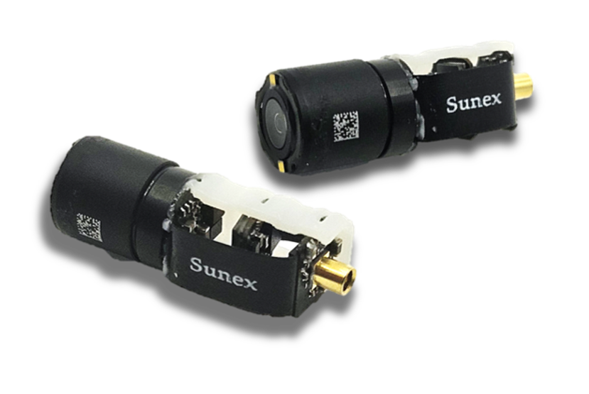
A lens is only as good as its position relative to the sensor. In a conventional assembly, small errors add up: element decenter inside the barrel, thread play, holder tilt, die placement on the package, and board flatness. Each one is small. Together, they tilt and shift the image plane.
That matters more than ever because pixels keep shrinking. As a rough guide, depth of focus is about ±2 x f-number x pixel pitch. For an F/2.0 lens on a 1.4 µm sensor, that’s only about ±6 µm. Typical assembly tolerances can easily use up that budget on one side of the image.
Active alignment solves this by aligning the lens to the live sensor rather than to mechanical datums:
- The sensor is powered and images a test target through the lens.
- A precision stage moves the lens in up to six axes (X, Y, Z, tip, tilt, and rotation).
- Software measures sharpness (MTF or SFR) across the field and finds the best position for center and corners together.
- Adhesive is cured in that position, locking the alignment permanently.
For medical devices, the benefits go beyond sharpness. There is no focus thread that can loosen under vibration or reprocessing, the module can be fully sealed, the optical center lands where the software expects it, and unit-to-unit consistency improves. That consistency simplifies calibration in diagnostic readers and stereo matching in DXM-based systems.
We’ve built camera modules around our own lenses for years, from optical design through active alignment and final test, so the lens and the module are engineered as one optical path rather than handed off between suppliers.
When active alignment isn’t necessary
Active alignment adds equipment and cycle time. For lower-resolution sensors or slower apertures, a well-designed threaded focus with a mechanical lock is often perfectly adequate, and we’ll recommend that when it fits.
What should you bring to your first conversation with a Sunex engineer?
The fastest programs we see start with a short, honest list of requirements, even if half of it is still marked “TBD.” These details let us suggest an off-the-shelf lens, a custom path, or both, in the first meeting:
- Sensor part number, pixel pitch, and format
- Field of view or object size, plus the working distance range
- Illumination and wavelength bands (visible, NIR, fluorescence)
- Mechanical envelope: diameter, length, and mounting
- Environment and reprocessing method (autoclave, low-temperature sterilization, disinfectant wipes, single use)
- Device classification and who holds design control
- Image quality targets (for example MTF at a given frequency, distortion, relative illumination)
- Expected volumes and timeline
Don’t have all of it yet? That’s normal. Bring what you have.
One more time, because it matters: the applications and lenses in this article are examples. Your device has its own requirements, and the best solution comes out of a detailed conversation with our engineers, not out of a product list.
Next Steps
→ Talk to a Sunex optical engineer about your medical imaging requirements: sunex.com/contact/
→ Browse the Sunex lens portfolio and order samples for your feasibility build: sunex.com/products/
→ Watch our YouTube Knowledge Library on MTF, distortion, and active alignment: youtube.com/@sunexinc
→ Read more articles in the Sunex Knowledge Center: sunex.com/support/knowledge-center
Still have questions about medical imaging optics?
Can an off-the-shelf lens be used in a regulated medical device?
Yes, in many cases. The device manufacturer qualifies the lens under its own design controls, the same way it qualifies other purchased components. What matters is that the lens meets the verified requirements and that lifecycle and change-notification terms are agreed with the supplier early in the program.
When does a custom lens design make sense for a medical program?
A custom design makes sense when an off-the-shelf lens cannot meet the mechanical envelope, the working distance, the spectral bands, or the image quality target, or when production volume is high enough that a design optimized for cost lowers the total unit price. Many programs start with an off-the-shelf lens for feasibility and move to a custom design for production.
Does Sunex offer off-the-shelf lenses small enough for endoscopes?
The smallest threaded off-the-shelf lenses use M7 and M8 mounts, which suit larger-diameter scopes, visualization handpieces, and proof-of-concept rigs. Distal tips smaller than roughly 4 mm require a custom miniature design.
What is active alignment, and why does it matter for medical camera modules?
Active alignment positions the lens relative to a powered, imaging sensor in up to six axes, measures sharpness across the field, and cures the adhesive at the best position. It compensates for assembly tolerances, improves corner-to-corner sharpness and unit-to-unit consistency, and allows sealed modules without focus threads.
How does DXM support 3D surgical visualization?
DXM projects two optical views onto a single image sensor, creating an inherently synchronized stereo pair in one compact module. It is best suited to short working distances, because the stereo baseline is limited by the sensor width.
Do veterinary devices need different optics than human-medical devices?
The optical physics is largely the same, but veterinary devices usually face lower price points, a wider range of patient sizes, and harsher use conditions. In the US they also do not require 510(k) or premarket approval submissions, which often favors off-the-shelf or derivative lens designs.